Peripheral Artery Disease Imaging: Assessing perfusion within the foot
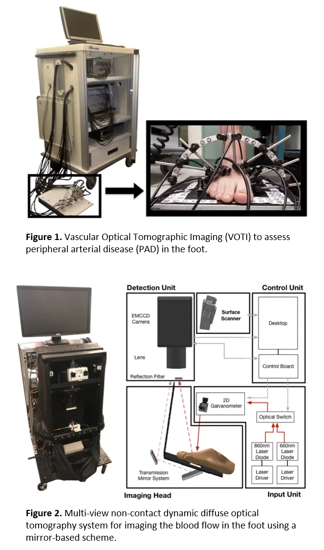
Peripheral artery disease (PAD) affects approximately 12 million people in the US and is considered the leading cause of low-extremity amputations, responsible for over 70,000 amputations annually. PAD is caused by an accumulation of plaque in the vessels of the lower extremities, which leads to stenosis and reduction in blood flow. If not properly treated, PAD can lead to critical limb ischemia with consequent cells death, ulcers, and gangrene. The disease affects mostly the elderly, diabetics, and patients with kidney problems. Early symptoms include claudication (pain while walking), leg numbness, or weakness and coldness in the lower leg or foot. To overcome some of these limitations, we have developed a vascular optical tomography imaging (VOTI) system, which measures transmitted and reflected light intensities and allows to estimate hemoglobin distribution inside the feet. In addition, our group has also developed a novel multi-view non-contact dynamic diffuse optical tomographic imaging system for the clinical evaluation of vasculature in the lower extremities by simultaneously obtaining and reconstructing diffusely reflected and transmitted light using a system of mirrors and a single CCD camera.